
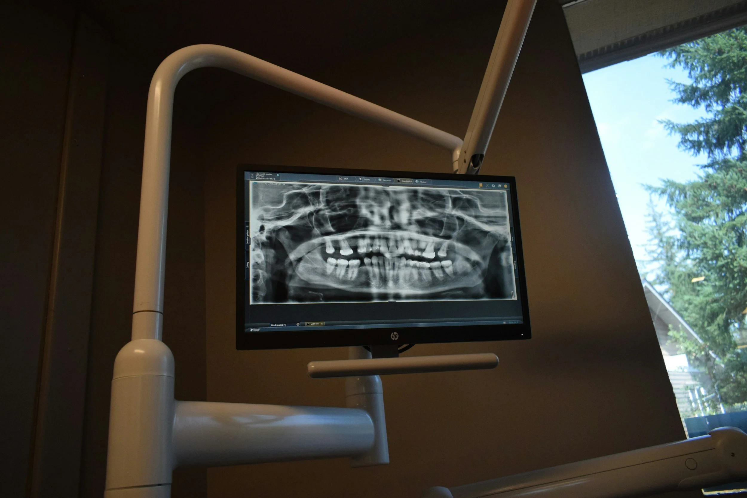
You sit down for your dental appointment, and before long someone asks a familiar question: "When was the last time you had X-rays?"
For many patients, dental X-rays are simply another part of visiting the dentist. Others understandably wonder why they are necessary, what dentists are actually looking for, and whether being exposed to radiation is something they should worry about.
Those are good questions.
Dental X-rays, also called dental radiographs, allow your dentist to see areas of your mouth that cannot be adequately evaluated with the naked eye. The important thing to understand is that X-rays are a diagnostic tool, not simply a box that needs to be checked at every appointment.
Here is what dental patients should know.
What Do Dental X-Rays Actually Show?
Even a very thorough visual dental examination has limitations.
Your dentist can examine the visible surfaces of your teeth and gums, but portions of each tooth are hidden below the gumline or pressed against neighboring teeth.
X-rays provide additional information about those areas.
Depending on the type of image being taken, dental X-rays can help identify or evaluate:
Cavities between teeth
Decay beneath or around existing restorations
The roots of teeth
Bone surrounding the teeth
Tooth infections and abscesses
Impacted or unerupted teeth
Periodontal bone loss
Development of children's permanent teeth
Previous dental work
Areas being evaluated for certain restorative procedures
Dental implant treatment planning
Think of the visual examination and dental X-rays as two different sources of information. One does not completely replace the other.
Why Can't a Dentist See a Cavity Without an X-Ray?
Some cavities are easy to identify because they develop on surfaces that are clearly visible.
Others are hidden.
One common location is between neighboring teeth. Because those surfaces touch or nearly touch one another, your dentist may not be able to directly see early decay developing there.
Certain dental X-rays can reveal changes in the tooth structure that would otherwise remain hidden.
This matters because a cavity that is identified relatively early may require less extensive treatment than one discovered after decay has progressed deeper into the tooth.
Are Dental X-Rays Safe?
Dental imaging does involve ionizing radiation, so patients are right to ask questions about safety.
Modern dental imaging is designed to obtain the diagnostic information a dentist needs while limiting unnecessary radiation exposure. Dental radiography represents a relatively small portion of a person's overall radiation exposure.
The guiding principle is straightforward: X-rays should be taken when there is a clinical reason for obtaining them, and exposure should be kept as low as reasonably achievable.
Advances in digital radiography have also made it possible to capture useful dental images efficiently while reducing many of the limitations associated with older film-based systems.
If you have questions or concerns about a recommended X-ray, ask your dentist what they are looking for and how the image may influence your treatment.
How Often Do You Need Dental X-Rays?
There is no universal answer.
You may have heard that everyone needs dental X-rays every six months, every year, or at some other fixed interval. Modern recommendations take a more individualized approach.
How frequently X-rays are appropriate can depend on factors such as:
Your age
Your dental history
Your current oral health
Whether you have active tooth decay
Your risk of developing cavities
Gum health
Previous dental restorations
New symptoms
Whether you are a new or established patient
The type of treatment being considered
Someone with an extensive history of dental decay may need imaging more frequently than a patient who has had healthy teeth and a very low cavity risk for many years.
Similarly, a patient experiencing unexplained tooth pain may need an X-ray even if their last images were taken relatively recently.
The goal is not to take images because a certain date appeared on the calendar. The goal is to obtain useful diagnostic information when that information can improve patient care.
What Are the Different Types of Dental X-Rays?
Not every dental X-ray is the same.
Bitewing X-Rays
Bitewings commonly show the crowns of upper and lower teeth in the same area of the mouth.
They are especially useful for examining spaces between teeth and identifying certain types of tooth decay that may not be visible during a normal examination.
Periapical X-Rays
A periapical image typically captures the entire tooth, including the crown, root, and surrounding bone.
These images can be particularly valuable when a dentist needs to evaluate a specific tooth, its root, or the tissues around it.
Panoramic X-Rays
A panoramic X-ray provides a broad view of the teeth, jaws, and surrounding structures.
Rather than focusing on just a few individual teeth, it gives the dentist a larger overview of the mouth and jaw.
3D Dental Imaging
Some situations may call for more advanced three-dimensional imaging, such as cone beam computed tomography, commonly called CBCT.
CBCT can provide highly detailed information useful for certain procedures, including some dental implant evaluations and other complex treatment planning.
Because 3D imaging involves different considerations than routine two-dimensional dental X-rays, it is generally used when the additional information is clinically valuable.
What If You Had X-Rays Taken at Another Dentist?
Tell your new dental office.
In many situations, existing dental records and X-rays can be transferred from one provider to another. If appropriate recent images are available and provide the necessary diagnostic information, repeating those same images may not be necessary.
Providing your dental history also gives your new dentist valuable context about previous treatments and changes in your oral health over time.
If you are changing dentists, asking your previous office to transfer your dental records can make the transition easier.
Dental X-Rays Help Dentists Find Problems You Cannot See
A tooth does not have to hurt for something to be happening below the surface.
That is one of the most useful things about dental imaging. X-rays can help dentists investigate areas they simply cannot see during a visual examination and can sometimes identify problems before they become obvious to the patient.
At the same time, modern dentistry does not require taking every possible image at every appointment. Your age, dental history, symptoms, disease risk, previous images, and treatment needs all help determine which X-rays are appropriate.
Lake Harbor Dentistry provides comprehensive family and restorative dental care in Boise, Idaho. If it has been a while since your last dental examination, or if you are experiencing tooth pain, sensitivity, swelling, or another unexplained change, an examination can help determine whether dental imaging would provide useful answers.
And if you are ever curious about an X-ray your dentist recommends, ask. Understanding what your dental team is looking for makes it much easier to be an active participant in your own oral health.
